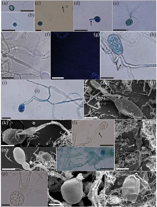
Capellomyces foraminis Hanafy, Vikram B. Lanjekar, Prashant K. Dhakephalkar, T.M. Callaghan, Dagar, G.W. Griff., Elshahed & N.H. Youssef, sp. nov.
MycoBank number: MB 830740; Index Fungorum number: IF 830740; Facesoffungi number: FoF;
Typification: The holotype (FIG. 4n) was derived from the following: USA. TEXAS: Val Verde County, 29.369°N, 100.829°W, ~300 m asl, 3-d-old culture of isolate BGB-11, originally isolated from freshly deposited feces of a female Boer goat (Capra aegagrus), Apr 2018, Radwa Hanafy. Ex-type strain: BGB-11. GenBank: MK881975 (D1–D2 28S rDNA).
Etymology: The species epithet “foraminis” refers to the wide apical pore at the top of the sporangia through which zoospores are discharged.
Obligate anaerobic fungus that produces spherical monoflagellate zoospores. Biflagellate zoospores are occasionally observed. Zoospores start to encyst after shedding their flagella. Zoospore cyst germinates, producing germ tube that subsequently branches into monocentric thalli with filamentous anucleate rhizoidal systems. Endogenous and exogenous sporangia are produced. Endogenous sporangia are ellipsoidal or ovoid. Exogenous sporangia are formed at the end of unbranched sporangiophores (ranging from 20 to 150 μm). Some of the sporangiophores exhibit subsporangial swellings. Exogenous sporangia are ovoid, ellipsoidal with a single constriction, and globose. Zoospores are liberated through a wide apical pore at the top of the sporangia followed by sporangial wall collapse. Colonies are small (0.1–0.5 mm diam), circular, and brown, with dark center of sporangia structures on agar. Produces thin fungal biofilm in liquid media. The clade is defined by the sequences MK882007–MK882009 (ITS1) and MK881975 (D1–D2 28S rDNA).
Additional specimens examined: USA. TEXAS: Val Verde County, 29.369°N, 100.829°W, ~300 m asl, 3-d-old culture of isolates BGB-2, BGC-12, BGS-11, and BGS-12, originally isolated from freshly deposited feces of a female Boer goat (Capra aegagrus), Apr 2018, Radwa Hanafy. GenBank (D1–D2 28S rDNA): BGB-2 (MK881974), BGC-12 (MK881976), BGS-11 (MK881977), and BGS-12 (MK881978).

Figure 4. Microscopic features of Capellomyces foraminis (clade 3, Boer goat) strain BGB-11 (W-TX). Light (a–i, l, o, p) and scanning electron (j, k, m, n, q, r) micrographs. Light microscopy images were examined after staining with lactophenol cotton blue (a–e, h, i, o), as well as following staining of nuclei with DAPI (f). g. Overlay image. a. A monoflagellate zoospore. b. A biflagellate zoospore. c. Zoospore cyst; arrow points to the shed flagellum. d. Germinating zoospore cyst producing a germ tube (arrow). e. Rhizoidal system development. f–g. Monocentric thalli with nuclei occurring in sporangia, not in rhizoids or sporangiophores. h–i. Endogenous sporangial development: h. Ellipsoidal sporangium with two main rhizoidal systems (arrows). i. Ovoid sporangium with single rhizoidal system. j–q. Exogenous sporangial development: j. Exogeous sporangium with a short sporangiophore (Sp); note the empty zoospore cyst (Zc). k. Ovoid sporangium with a long sporangiophore. l. Ovoid sporangium on a long sporangiophore ending with subsporangial swelling (arrow). m. Collapsed empty sporangium on a long sporangiophore ending with subsporangial swelling (arrow). n. Constricted ellipsoidal sporangium. o–p. Globose sporangia. q. Zoospores are released through apical pore. r. An empty sporangium following zoospores release. Sp = sporangiophore; Zc = zoospore cyst. Bars: a–j, l, o, p = 20 μm; k, m, n, q, r = 50 μm.
